ISSN Number
ISSN 2771-019X-

-

Impact Factor
1.2*
ISSN Number
ISSN 2771-019X
Impact Factor
1.2*Department of Operative Dentistry, School of Dentistry, National and Kapodistrian University of Athens, Greece.
Department of Operative Dentistry, School of Dentistry, National and Kapodistrian University of Athens, Greece.
Tel: +306977579857, Fax: +302107222860;
Email: sthomaidisgr@yahoo.com
Received : Jan 04, 2025,
Accepted : Feb 12, 2025
Published : Feb 19, 2025,
Archived : www.jclinmedcasereports.com
A porcelain fused to metal partial coverage overcasting was made, in order to repair a broken veneering ceramic on an existing multi-unit porcelain fused to metal bridge. This crown served as an abutment for a removable partial denture. Clinical and laboratory procedures of the fabrication of the porcelain fused to metal overcasting, as well as the removable partial denture, are described. Critical points affecting survival of the overcasting, as well as the existing bridge, are elaborated.
Keywords: Metal ceramic failure; Metal ceramic chipping; Metal ceramic repair; Overcasting.
Copy right Statement: Content published in the journal follows Creative Commons Attribution License (http://creativecommons.org/licenses/by/4.0). © Thomaidis S (2025).
Journal: The Journal of Clinical and Medical Images, Case Reports (JCMICR) is a fantastic resource for keeping up with the latest clinical advancements and for publishing case reports and clinical images related to a variety of medical illnesses.
Porcelain fused to metal failure due to porcelain chipping, extensive veneering ceramic fracture is reported to occur between 2,3% and 8% in a 5-year period, and is attributed to occlusal forces, and non-properly supported porcelain by the metal framework [1-3]. When veneering porcelain fracture occurs in a part of a metal ceramic bridge, different treatment plans can be performed, including removal of the existing bridge and fabrication of a new one, and intraoral repair with composite resin [4]. The repair with composite resin, can be done, after preparing/roughening the porcelain and metal of area to be restored with a diamond bur or/and sandblasting with an intraoral microetcher, followed by etching the porcelain with hydrofluoric acid under rubber dam isolation, placement of a primer, opaquer, if needed, and finally composite resin, that would be finished and polished. The repair of a porcelain fused to metal crown or bridge presenting small to moderate chipping or fracture, is not so effective in terms of survival time, and is considered as a temporary measure [5]. An indirect repair technique has been previously reported [6].
The aim of this study was to describe the clinical and laboratory stages of the fabrication of a partial coverage porcelain fused to metal superstructure, and a removable partial denture, in order to restore the existing multi-unit porcelain fused to metal bridge, and edentulous area, in terms of function, as well as esthetics. Factors affecting success will be discussed.
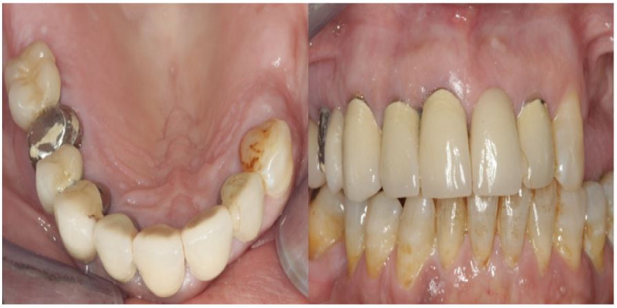
A 60-year-old female patient, with free medical history, presented at the office. The patient was treated for moderate chronic periodontitis by a periodontist, and was on 6-month recall. The patient had a long span maxillary porcelain fused to metal bridge, where teeth no 4,6,7,8, and 10 served as abutments, teeth no 5 and 9 were restored as pontics, and tooth no 3 was a cantilever (Figure 1a & b). Tooth no 11 did not have any restorations. Root canal therapy had been performed on teeth no 4, 6, 7, and 8, without any sigh or symptoms of inflammation. None of the teeth, having root canal therapy, had any kind of post and core. In the mandible teeth no 20, 34 were missing, as well as the wisdom teeth. The patient had no intention to restore tooth no 34.
The patient was informed about the different treatment plans, including a new maxillary multi-unit bridge, including post and cores if needed, a repair with composite resin, and the partial coverage metal ceramic overcasting, which was selected. Abutment tooth no 4 presented extensive ceramic delamination, implying that the metal framework oxidation was probably not properly performed.
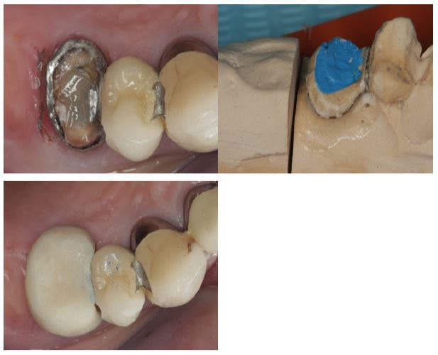
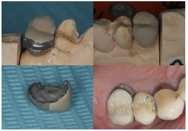
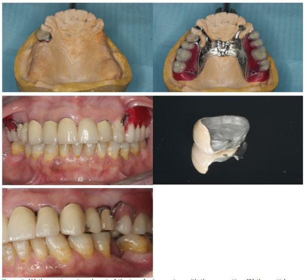
Initially, tooth no 3, which served as a cantilever, was sectioned and removed. Tooth no 4 was prepared, removing 1,5 mm to 2 mm occlusally, in order to provide enough space for the metal ceramic overcasting. The metal framework was prepared, removing all of the porcelain left. The metal framework of tooth no 4, was prepared circumferentially more than 180O, but in the least possible depth, in order mainly to create a path of insertion with minimum convergence, not extending in the connector area, in order not to jeopardize the stability of the connector, and as a consequence the survival of the existing bridge. Therefore, the connector area was left unprepared, as much as possible. Margin preparation was performed with a chamfer diamond bur, and was done as minimally as possible (Figure 2a). The margins of the existing abutment were left unprepared, leaving an as thin as possible metal collar, that was polished with a fine diamond bur in an airotor handpiece, followed by the use of rubber polishing tips with a micromotor handpiece. A double mix heavy body/light body poly-vinyl-siloxane impression was made, sent to the lab, and a cast with removable dies was poured in extra hard stone (Figure 2b). A provisional restoration was made out of methyl-methacrylate (Figure 2c). A wax pattern was made and cast in base metal alloy, incorporating a lingual ledge to accommodate the bracing clasp arm of the removable partial denture, and act partly as a rest. A metal try-in was performed. Afterwards porcelain was fired and a partial coverage metal ceramic overcasting was made (Figure 3a, b & c), a bisque bake try-in was performed (Figure 3d). The mesial lingual area of teeth no 7 and 11, as well as the distal lingual area of teeth no 6 and 10 were prepared, in order to achieve a positive rest area. In teeth no 6 and 10, it acted as an indirect retainer. A transfer (pick-up) double mix heavy body/light body poly-vinyl-siloxane impression was made, in order to pour a new cast in extra hard stone, with a fixed acrylic die, and the metal ceramic overcasting on it (Figure 4a). A Removable Partial Denture (RPD) was designed upon this stone model with the use of a surveyor, and the RPD metal framework was ordered. The RPD metal framework consisted of a U-shaped palatal major connector. Tooth no 11 received an RPI design clasp, and tooth no 4 an I bar retentive arm. Teeth were arranged, an acrylic tooth try-in was performed (Figure 4b & c), and the RPD was sent to the lab, for flasking, the wax elimination, processing in pink fibered acrylic resin, finishing and polishing. The partial coverage porcelain fused to metal overcasting was then glazed (Figure 4d) The removable partial denture was delivered, after relieving pressure point areas, and adjusting occlusion (Figure 4e). The crown was then cemented with the RPD seated in place upon it. The preparation of no 4, as well as the interior surface of the overcasting, were sandblasted with the use of an intraoral sandblasting instrument and 50 μm Aluminum oxide particles. The overcasting was cemented with a dual-cure resin luting cement.
Different treatment modalities could be used, such as the repair with composite resin, and fabrication of a new porcelain fused to metal bridge from #4, all the way to #11, along with a removable partial denture.
The repair with composite resin, after etching chipped porcelain with hydrofluoric acid, followed by silane application, can lead to an acceptable repair [4]. However, when the fracture occurs with metal exposure, the repair is more problematic, due to low bond strength of the metal framework, with the composite resin [5]. In the case presented, abutment tooth no 4 demonstrated a complete delamination, including the occlusal surface, a stress bearing area, and this tooth would also serve as an abutment for a removable partial denture. Therefore, the repair with composite resin was considered a temporary solution, with an even more limited life, and therefore not recommended.
From a meta-analysis, Sailer et al [1] estimated the survival rate of metal-ceramic single crowns being 94.7% after 5 years. In another meta-analysis 86.7% of the metal‐ceramic single crowns experienced no biological or technical complications over the 5-year observation period [3]. Pjetursson et al [2] estimated a 94.4% 5-year survival rate of metal-ceramic multiple unit fixed partial dentures. A new metal ceramic bridge would be the best choice of treatment in terms of longevity and esthetics, but the main disadvantage is cost, followed by multiple appointments, as well as discomfort for the patient. Removing the existing multi-unit bridge could also result in damaging some of the abutment teeth.
The survival of this type of partial coverage restoration depends primarily on the survival of the existing bridge. Therefore, care should be taken during preparation of the abutment tooth, to remove the least metal possible from its framework, and primarily not to weaken the connector. The metal framework should be left intact as much as possible circumferentially, with minimal preparation lingually, where esthetics are not important. The pre-existing bridge framework would be better not to be totally eliminated occlusally, in order to preserve the bridges resistance to deformation. Nevertheless, a minimum of 1,5 mm occlusal space is needed for the over casting. When the framework of the existing bridge is prepared, there is no information about its metal framework thickness, and as a consequence it is better to perform minimum metal preparation, especially in the case it was initially made with minimal inadequate preparation. The aim of the preparation of the existing metal framework is to keep as much as possible of the metal framework thickness, in order to preserve the stability of the existing bridge, and just to create a path of insertion. In this case, the initial preparation of tooth no 4 was not performed properly, it was underprepared. The overcasting would anyways present slightly over contoured, since at least 0,4 mm of additional space would be needed, ie 0,1 mm for the existing cement, and 0,3 mm for the existing metal framework, considering that the overcasting needs at least a space of 1,3 mm, ie 0,3 mm for the metal framework, 0,2 mm for the opaque layer, and 0,7 mm for dentine and enamel porcelain [7,8], as well as 0,1 mm for the cement. In this case the overcasting was over contoured, since the initial bridge was so, as well.
The longevity of the overcasting is enhanced by increasing its retention. This restoration has limited retention compared to a full coverage crown. Retention can be increased, by increasing roughness, surface, and minimizing the convergence angle of the walls as close to 2-3o as possible [9]. Longer preparation results in increased retention. Surface can be also augmented, by sandblasting, with an intraoral micro etcher, the prepared pre-existing metal framework, as well as the interior surface of the overcasting, and thus lead to increased bond strength [10]. Resin luting cement can also lead to higher dislodgement forces compared to traditional cements, such as zinc phosphate or glass ionomer, and therefore it is recommended [11]. Retention can be increased by preparation of longitudinal grooves, such as the ones placed buccally and lingually to the connector [9]. In case of groove preparation, care should be taken not to weaken substantially the existing metal framework.
The load exerted by the RPD on the overcasting should be minimized as possible by having an accurate impression, and placing stress braking claps, such as the RPI [12,13]. The clasp placed on tooth no 4 was RPI-type, incorporating a buccal I bar designed mesially of the long axis of the tooth, which can provide stress-braking action. Whatsoever the lingual bracing arm set on a ledge on the crown, can act as a positive rest, but can exert higher forces, than a pure RPI clasp, on the abutment tooth. This partial coverage metal ceramic restoration is cost and time effective, since the existing prosthesis can be salvaged, and requires fewer appointments and discomfort for the patient. However, to prevent further complications such as crown loosening or fracture of the existing bridge, this method should be used cautiously. It is recommended that various efforts be made to eliminate the possibility of pre-existing bridge fracture or overcasting loosening, such as overpreparation of the existing bridge, large convergence angle of the preparation, inappropriate occlusion, premature contacts, parafunction, and misfit of the overcasting. The expected survival time of a partial coverage metal ceramic crown is probably shorter than that of a full metal ceramic crown, due to limited retention.
A partial coverage porcelain fused to metal overcasting can be a viable restoration of an extensively fractured, chipped, or delaminated multi-unit metal ceramic bridge, with the advantage of being more cost and time effective.
Poster presentation: Presented at the 40th Panhellenic Dental Congress (6-8 October 2022) in Athens, Greece as a poster.Conflict of interest: The author of this article is certifying that he has no proprietary, financial, or other personal interest of any nature or kind in any product, service, and/or company that is presented in this article.
Funding: No funding was received for this manuscript.
