ISSN Number
ISSN 2771-019X-

-

Impact Factor
1.2*
ISSN Number
ISSN 2771-019X
Impact Factor
1.2*1Anicura-Istituto Veterinario di Novara, S.P.9 Granozzo con Monticello, Italy.
2Centro Veterinario Cattolica, Via Toscana 11, Italy.
Anicura-Istituto Veterinario di Novara, S.P.9 Granozzo con Monticello, Italy.
Email: info@giordanazanna.it
Received : Feb 03, 2025,
Accepted : Mar 07, 2025
Published : Mar 14, 2025,
Archived : www.jclinmedcasereports.com
Three unrelated indoor/outdoor domestic short hair adult cats, presenting with clinical signs of pruritus of varying degrees, were diagnosed with trombiculiasis through the in vivo dermoscopic visualization of Trombiculidae larvae. Although the parasites were also demonstrated via microscopic examination of both adhesive tape impressions and skin scrapings from the affected skin, for the first time, dermoscopy provided a non-invasive, readily available, and useful tool for the early detection of trombiculosis in cats.
Keywords: Dermoscopy; Chigger mites; Cats.
Copy right Statement: Content published in the journal follows Creative Commons Attribution License (http://creativecommons.org/licenses/by/4.0). © Zanna G (2025).
Journal: The Journal of Clinical and Medical Images, Case Reports (JCMICR) is a fantastic resource for keeping up with the latest clinical advancements and for publishing case reports and clinical images related to a variety of medical illnesses.
In humans, there is emerging evidence that dermoscopy is a valuable aid to the available tools for diagnosing several parasitic skin diseases (e.g. scabies, phthiriasis, pediculosis, hookworm-related cutaneous larva migrans, tick infestations and myiasis) [1,2]. In 2006, Scanni and Bonifazi proposed a neologism to describe the dermoscopic findings in ectoparasitosis, leading to the introduction of the term entodermoscopy [3]. Recently, the usefulness of this technique has also been documented in diagnosing trombiculosis, an epizoonosis that occurs worldwide, offering the opportunity to rapidly identify the parasite [4,5].
This challenge has also been exemplified in veterinary dermatology in the diagnosis of canine scabies. The authors described the first two cases of sarcoptic mange in which dermoscopy was used for in vivo visualization of the adult mites, demonstrating as this technique may be considered a rapid, point-of care test in cases of heavy parasitic load [6].
To further support the impact that this tool may have on the identification of the parasites on the skin surface, we document the dermoscopic detection of chigger mites in three cats naturally infested with trombiculosis.
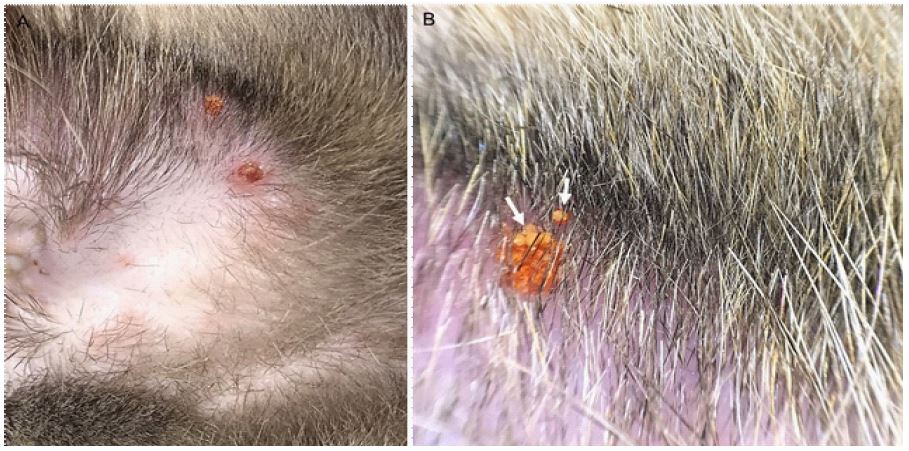
Case 1: A 1-year-old neutered male domestic long-haired cat was presented with a 10-day history of pruritus on the head, and crusts on the chin and auricular pinna. The cat lived both indoors and outdoors with other cats, was fed commercial food, not regularly vaccinated, and treated monthly against ectoparasites with lotilanerTM tablets (Credelio, Elanco). Dermatological examination revealed scales, crusts, and orange-red dots on the ear tips and inner pinna folds (Figure 1A). Similar lesions were also observed on the chin, the external side of the left eye, and the left labial commissure. A dermoscopic examination was conducted using a handheld light dermoscope (Handyscope for iPhone SE, Fotofinder Systems GmbH; Bad Birnbach, Germany) connected to a smartphone (iPhone SE, Apple; Cupertino, CA, USA). Alcohol (Kodan® spray, Schulke & Mayr, Vienna, Austria) was applied to the skin as a contact solution to enhance the visibility of surface and subsurface microscopic features and minimize light reflection. Images at ×10 magnification were obtained and stored through a dedicated iPhone app (Handyscope3® app - version 3.0.6, FotoFinder Systems GmbH Bad Birnbach, Germany). Aggregates of reddish, round corpuscles, resembling Neotrombicula larvae, were clearly observed among the crusts and scales (Figure 1B). Adhesive tape impressions and multiple skin scrapes also confirmed the diagnosis of parasite infestation. Fipronil pump spray 0.25% (Frontline, Boehringer Ingelheim) was applied to the affected areas every 7 days whereas fipronil spot on (Frontline, Boehringer Ingelheim) was administered once on the dorsal neck. After four weeks, there were no longer any signs of infestation, and the lesions had completely healed.
Case 2: A 4-year-old spayed female domestic short-hair cat was presented with bilateral orange-red spots on the ear pinnae. The cat was otherwise healthy, showing only mild facial pruritus. The cat lived both indoors and outdoors, was fed commercial food and was regularly vaccinated, but had not been regularly treated for ectoparasites.
Dermatological examination revealed orange-red clumps on both auricular pinnae, inner pinnae folds, and the skin of the temples bilaterally (Figure 2A). Dermoscopic examination was performed as reported in case 2, and small rounded corpuscles, within the crusts and scales referable to Neotrombicula autumnalis mites, were detected (Figure 2B). Adhesive tape impressions and skin scrapes confirmed the parasitic infestation.
Fipronil spot on (Frontline, Boehringer Ingelheim) was administered once on the dorsal neck, and one spray of fipronil pump-spray 0.25% (Frontline, Boehringer Ingelheim) was applied weekly to the pinnae. After 10 days a significant improvement was already detected with no signs of infestation after one month.
Case 3: A 2.5-year-old neutered male domestic shorthair cat, with access to both indoor and outdoor environments, presented with a one-month history of a patch of hair loss on his chin region associated with mild pruritus. The patient received irregular ectoparasites prophylaxis, was regularly vaccinated and fed commercial food.
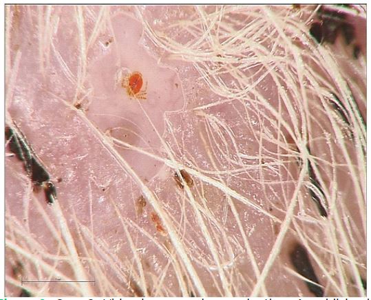
Dermatological examination revealed alopecia, yellow-brown scaling, and orange dots on the chin region. Dermoscopic examination was performed using a videodermoscope (Fotofinder Systems GmbH; Bad Birnbach, Germany) that allowed for magnification up to ×70. Alcohol (Kodan® spray, Schulke & Mayr, Vienna, Austria) was applied to the skin as an interface solution to enhance the observation of surface and subsurface microscopic features and reduce light reflection. At ×70 magnification, reddish mites attached to the skin on the patient’s chin were clearly visible (Figure 3). Stored images revealed six-legged larvae consistent with Neotrombicula autumnalis. The larvae were measured using the integrated software in the videodermoscope, with a mean length of 0.25 mm. Acetate tape impression tests from the chin area confirmed the presence of the suspected mites.
Every week the patient was treated with one spray of fipronil pump-spray 0.25% (Frontline, Boehringer Ingelheim Pharma GmbH Germany) on the chin region whereas fipronil spot on (Frontline, Boehringer Ingelheim Pharma GmbH Germany) was applied once between the shoulders. No signs of infestation were observed after one month of treatment.

The dermoscopic findings presented herein offer significant insights into the clinical implications of the presence of the parasite on the skin of cats affected by trombiculosis.
In general, the larval stage of trombiculid mites, also known as “chiggers” or “harvest mites,” represents the only stage capable of parasitizing a wide variety of warm-blooded vertebrates worldwide. These mites are ubiquitous, and depending on favorable climatic conditions, natural infestation typically occurs during warmer and wet late summer or autumn months, with the host in the outdoor environment being attacked near points of contact with infested vegetation [7,8]. In cats with access to rural areas, characteristic orange-red spots or granules are commonly found at the marginal pouch at the base of the pinnae (Henry’s pocket), chin, or interdigital spaces, likely due to their exploratory behavior that allows the larvae to come into direct contact with them [9]. Although the larvae may be visible to the naked eye as orange specks, a definitive diagnosis requires microscopic examination of superficial skin scrapings or adhesive tape impressions [9,10].
To date, a wide range of anti-flea products have been demonstrated to effectively protect against harvest mites even if they are not specifically registered for trombiculid mites. Among these there is fipronil in both spray and spot-on formulations, intended as a safe and practical treatment for eradicating localized infestations of Neotrombicula species in cats [11,12]. However, the spray must be applied thoroughly, and some cases may require additional topical treatments presumably due to the more fastidious grooming habits of felines [12]. In this case series, the combination of fipronil spray at 7-day intervals and monthly fipronil spot-on applications was demonstrated to ensure direct efficacy against already attached parasites.
The focus of this report was the acquisition and storage of images using dermoscopy, thus representing a significant advancement in diagnostic methodology. This approach facilitated the timely identification and, through videodermoscopy equipped with specialized software, also a precise measurement of chigger mites. To the best of the authors’ knowledge, the length of the Trombicula larvae has never been described before using dermoscopy.
In general, handheld dermoscopes, the most widely used devices, provide low magnification. Nevertheless, their practicality, excellent optical resolution, and high image quality make them ideal for everyday clinical use. Conversely, videodermoscopes are more advantageous for research purposes due to their higher magnification, measurement capabilities, superior resolution, and calibration features [13]. All these characteristics taken together simplify the processes of image viewing, storage, analysis, and retrieval processes, thereby enhancing their versatility.
For the first time, Trombicula mites have been detected by dermoscopy in cats. This case series opens new horizons for the utility of this non-invasive skin imaging technique also in veterinary dermatology.
