ISSN Number
ISSN 2771-019X-

-

Impact Factor
1.2*
ISSN Number
ISSN 2771-019X
Impact Factor
1.2*Department of Radiology, Children’s Hospital, Rabat, Morocco.
Department of Radiology, Children’s Hospital, Rabat, Morocco.
Tel: +212 6 59290518
Email: salma.elkadiri.med@gmail.com
Received : Jul 09, 2025 ,
Accepted : Aug 21, 2025
Published : Aug 28, 2025,
Archived : www.jclinmedcasereports.com
A porcelain aorta is characterized by severe circumferential calcification of the ascending aorta and arch. This condition poses significant challenges for cardiac surgery due to the difficulty of clamping the aorta. This condition, primarily caused by atherosclerosis, is prevalent among the elderly and individuals with valve or coronary artery disease. Identifying it during preoperative imaging is critical for preventing intraoperative problems and guiding alternative surgical methods. This case describes the inadvertent detection of a porcelain aorta via chest computed tomography, underscoring the importance of a thorough preoperative examination.
Copy right Statement: Content published in the journal follows Creative Commons Attribution License (http://creativecommons.org/licenses/by/4.0). © Kadiri SE (2025)
Journal: The Journal of Clinical and Medical Images, Case Reports (JCMICR) is a fantastic resource for keeping up with the latest clinical advancements and for publishing case reports and clinical images related to a variety of medical illnesses.
A 56-year-old man was admitted for surgery to treat severe aortic insufficiency. He had a medical history of Behçet’s disease, hypertension, and chronic aortic regurgitation. An elective aortic valve replacement was planned for him. A chest computed tomography performed as part of the preoperative evaluation revealed a surprising amount of circumferential calcification of the ascending aorta, which is consistent with a porcelain aorta (Figure 1).
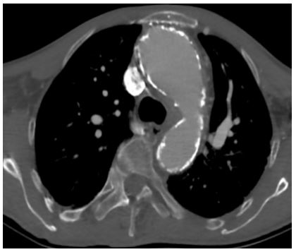
A porcelain aorta is defined as widespread, often circumferential, calcification of the thoracic aorta, primarily affecting the ascending aorta and the aortic arch. This disease poses a significant problem in cardiac surgery because clamping or manipulating a severely calcified aorta increases the risk of embolism, dissection, and hemorrhage, requiring alternative surgical methods.
Underlying causes include intimal atherosclerosis, linked to traditional cardiovascular risk factors, and medial calcification, which is typically age-related or associated with chronic inflammatory or metabolic illnesses. These processes may coexist, resulting in the widespread stiffening of the aortic wall.
Despite its importance in surgical planning, porcelain aorta lacks a clear definition and is not usually included in preoperative risk assessments. Its detection is thus mostly based on imaging.
Non-contrast chest computed tomography is the most effective imaging technique for detecting and characterizing aortic calcifications. It enables a precise assessment of location, extent, and circumferential involvement, which is critical for surgical risk categorization. Early detection of porcelain aorta allows the heart team to modify the operating strategy and investigate less invasive options such as off-pump surgery, transcatheter aortic valve replacement, or peripheral cannulation when traditional access is not possible.
