ISSN Number
ISSN 2771-019X-

-

Impact Factor
1.2*
ISSN Number
ISSN 2771-019X
Impact Factor
1.2*Department of Ophthalmology, The Royal London Hospital, Barts Health NHS Trust, London, UK.
Department of Ophthalmology, The Royal London Hospital, Barts Health NHS Trust, London, UK.
Email: neil.shah5@nhs.net
Received : Aug 16, 2025,
Accepted : Sep 02, 2025
Published : Sep 09, 2025,
Archived : www.jclinmedcasereports.com
Copy right Statement: Content published in the journal follows Creative Commons Attribution License (http://creativecommons.org/licenses/by/4.0). © Shah N (2025).
Journal: The Journal of Clinical and Medical Images, Case Reports (JCMICR) is a fantastic resource for keeping up with the latest clinical advancements and for publishing case reports and clinical images related to a variety of medical illnesses.
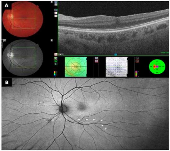
A 59-year-old man presented with vertical binocular diplopia following left vitrectomy Epiretinal Membrane (ERM) peeling, cryotherapy to a longstanding superior retinal tear and air tamponade. The unaided visual acuity OS was 6/9 (Snellen).
On post-operative examination, his retina was fully attached and ocular motility full. Optical Coherence Tomography (OCT) of the macula (A) showed no residual ERM with a good foveal contour. Fundus autofluorescence imaging (B) revealed inferior retinal displacement as the cause of his symptoms. The arrows (B) map areas of hyper-autofluorescence following the course of the pre-operative retinal vasculature.
The aetiology of retinal displacement in this case is unknown. Possible causes include retinal traction and displacement during the epiretinal membrane peel, which could include a localised macular detachment. Extension of subretinal fluid from the superior retinal tear may explain posture dependent displacement under air tamponade but this seems unlikely, since the displacement affected the inferior vascular arcade only and not the superior arcades. Management options include conservative with prismatic correction, macular relocation surgery or occlusion.
This case highlights an uncommon complication of ERM surgery and the importance of fundus autofluorescence as a diagnostic tool in post-operative diplopia. Given this complication, this case highlights the importance of careful pre-operative patient counselling with respect to the risks of harm from surgery.